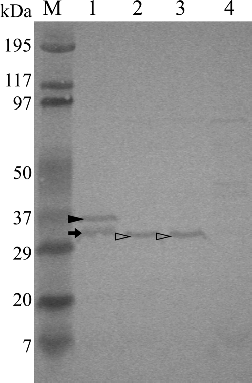
Fig 3.
Detection of PaCdtB of wild-type P. alcalifaciens by Western blotting. Whole-cell lysate of P. alcalifaciens was separated by SDS-PAGE (15%), transferred into a PVDF membrane, and probed with rabbit anti-rPaCdtB antiserum, followed by treatment with goat anti-rabbit IgG tagged with HRP as a secondary antibody. Color development was performed with 4CN-PLUS. Lanes: M, prestained SDS-PAGE broad-range marker (Bio-Rad);1, purified rPaCdtB; 2, bacterial lysate of AH-31 (P. alcalifaciens); 3, bacterial lysate of AS-1 (P. alcalifaciens); 4, bacterial lysate of E. coli strain BL21(DE3) carrying empty vector pET28a. The closed and open arrowheads and the arrow indicate rPaCdtB, PaCdtB, and degraded product of rPaCdtB, respectively. The experiment was carried out at least thrice.

