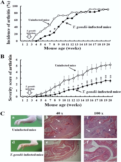
Fig 1.
Effects of T. gondii infection on spontaneous development of arthritis in IL-1Ra-deficient mice. (A) IL-1Ra-deficient BALB/c mice were p.o. infected with 10 T. gondii cysts at 2.5 weeks, and the development of arthritis in uninfected (open circles) and infected (closed circles) mice was examined weekly as described in Materials and Methods. Incidence (percentage) was calculated from 11 uninfected and 29 T. gondii-infected mice. (B) The severity scores of arthritis in uninfected (open circles) and infected (closed circles) mice were determined weekly as described in Materials and Methods. The average scores ± standard deviations (SD) were obtained from 11 uninfected and 29 T. gondii-infected mice. Statistical differences between uninfected and T. gondii-infected groups are shown as follows: #, P was <0.05 by 2-by-2 contingency χ2 test; *, P was <0.05; and **, P was <0.01 by Student's t test. (C) Histopathology of the ankle joints of uninfected (a to c) and T. gondii-infected (d to f) IL-1Ra-deficient BALB/c mice. (a) Swelling and redness of the ankle joint of an uninfected IL-1Ra-deficient BALB/c mouse. (b, c) Paraffin-embedded tissue sections of ankle joints from uninfected IL-1Ra-deficient mice were stained with hematoxylin/eosin, and microscopic views magnified at ×40 and ×100 are shown. Marked infiltration of inflammatory cells (arrows), hyperplasia of synovial membrane (stars), and erosive destruction of the bone (arrowheads) were observed. (d) Ankle joint of a T. gondii-infected IL-1Ra-deficient mouse. Swelling and redness of the ankle joint are not observed. (e to f) Microscopic observation of the ankle joint of a T. gondii-infected IL-1Ra-deficient mouse without infiltration of inflammatory cells.

