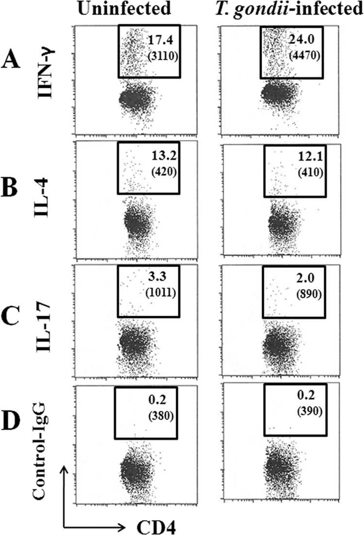
Fig 5.

Flow cytometric analyses at the chronic phase of infection. Spleen cells of T. gondii-infected IL-1Ra-deficient mice without the development of arthritis at 8 weeks p.i. were stained with those of age-matched uninfected mice for intracellular IFN-γ (A), IL-4 (B), or IL-17 (C) as described in Materials and Methods. (D) An isotype control was used for staining. Numbers shown in each square are the percentages of cells contained in gated CD4+ cells. The mean fluorescent intensity (MFI) is given in parentheses. Experiments were repeated three times with similar results.
