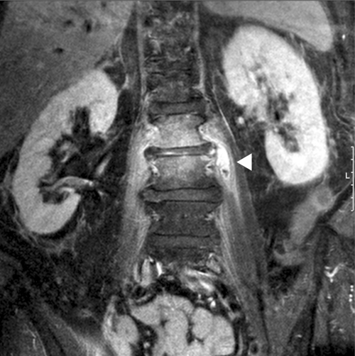
Fig 1.
Magnetic resonance imaging of the patient's spine. Signs of spondylodiscitis between lumbar bodies 2 and 3, with a reduction of lumbar disc height and diffuse enhancement caused by inflammation, are visible. Abscess formation (arrow) in the left psoas muscle with surrounding inflammation is shown. Coronary view, fat-suppressed T1 image after gadolinium application.

