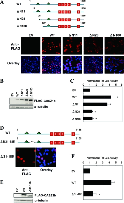
Fig 5.
The CASZ1b transcriptional activation domain is defined by aa 31 to 185. (A) Schematic of the full-length CASZ1b protein with the two NLSs (triangles) and the five ZFs (boxes) indicated. The three N-terminal deletions are also indicated. Colocalization of WT FLAG-CASZ1b and the three N-terminal variants with the nucleus is shown underneath. Chromatin is stained with DAPI. Magnification, ×63. (B) Western blot showing steady-state WT and variant CASZ1b protein levels 24 h after transfection. (C) Activation of the TH-luciferase construct by WT CASZ1b and the three N-terminal deletions 24 h after transfection. Two of the mutants lose complete function, while only ΔN11 retains function (*, P < 0.001). (D) Cellular localization of the ΔN31-185 CASZ1b mutant protein after staining with anti-FLAG antibody. Chromatin is stained with DAPI. Magnification, ×63. (E) Western blot showing steady-state WT and ΔN31-185 CASZ1b protein levels 24 h after transfection. (F) Activation of the TH-luciferase construct 24 h after transfection of 293T cells with WT CASZ1b and ΔN31-185 CASZ1b plasmids (*, P < 0.001).

