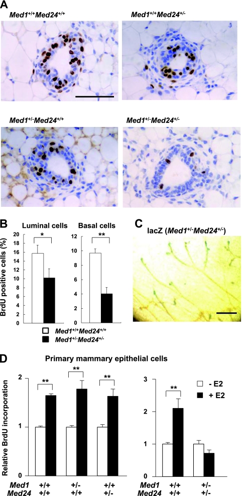
Fig 6.
Retarded DNA synthesis of Med1+/− Med24+/− mammary epithelial cells. (A) BrdU immunohistochemical staining of mammary inguinal gland sections. After 2 h of purging 8-week-old littermate virgin females with BrdU, BrdU-positive cells are visualized. Representative sections at terminal buds are shown. Scale bar, 50 μm. (B) Percentage of BrdU-positive mammary luminal and basal cells from wild-type and Med1+/− Med24+/− mice are shown (n = 10). The values represent means ± SE (*, P < 0.05; **, P < 0.01). (C) Whole-mount lacZ staining of 8-week-old Med1+/− Med24+/− mammary inguinal gland. Terminal buds are positively stained. Scale bar, 1 mm. (D) BrdU incorporation into primary mammary epithelial cells after E2 addition. The incorporation is comparable for singly Med1+/− or Med24+/− cells (left panel) but impaired in Med1+/− Med24+/− cells (right panel). Values (means ± SD from representative experiments performed in duplicate) are plotted as a fold increase against the value obtained without E2 (**, P < 0.01).

