Fig 4.
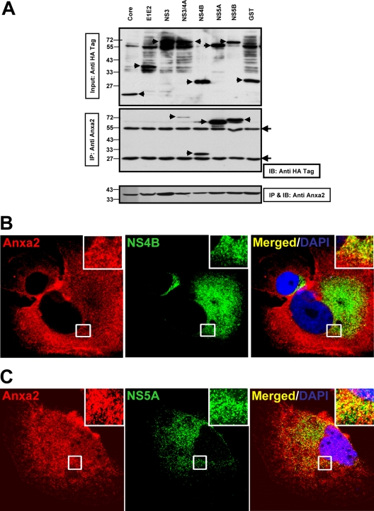
Anxa2 interacts with HCV nonstructural proteins. (A) Huh7.5 cells were transfected with plasmids encoding HA-tagged HCV NS proteins or GST. At 48 h posttransfection, cell lysates were immunoprecipitated (IP) using anti-Anxa2 monoclonal antibodies. The immunoprecipitates and 5% of the total cell lysates were subjected to 10% SDS-PAGE followed by Western blotting with anti-HA and anti-Anxa2 antibodies. (Upper panel) Anti-HA immunoblot showing the input proteins (arrows) (5% total). (Middle panel) Anti-HA immunoblot showing HCV NSP coimmunoprecipitated by anti-Anxa2 antibody. (Lower panel) Anxa2 protein immunoprecipitated by anti-Anxa2 antibody. Protein molecular mass markers are indicated on the left side. Filled arrowheads indicate the IgG heavy (55-kDa) and light (25-kDa) chains. (B and C) Colocalization of NS4B (B) and NS5A (C) with Anxa2 in HCV-JC1-infected Huh7.5 cells. Images shown were collected sequentially with a confocal laser-scanning microscope and merged to demonstrate colocalization. Enlargements of the sections, from different areas of the cells, indicated by the white squares are shown in the inset panels.

