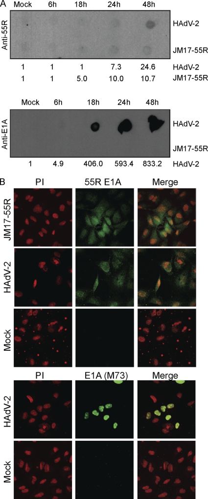
Fig 3.
Expression kinetics and subcellular localization of 55R E1A during HAdV-2 infection. Human A549 cells were infected with HAdV-2 or JM17-55R at an MOI of 10 or were mock infected. (A) Cells were collected at 6, 18, 24, or 48 hpi, and 5 μg of nonreduced lysate was blotted using anti-55R E1A polyclonal Abs or anti-E1A (M73). Numbers beneath blots are densitometry readings. (B) Subcellular localization of 55R E1A during infection was assessed at 48 hpi. Cells were stained with either anti-55R E1A polyclonal Abs or anti-E1A (M73), followed by Alexa Fluor 488. Nuclei were stained with propidium iodide (PI).

