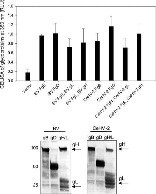
Fig 1.
Expression of gB, gD, and gH/L from BV and CeHV-2 on the cell surface by CELISA and in whole-cell lysates by Western blotting. The upper panel shows the CELISA for cell surface expression of the glycoproteins from BV and CeHV-2. CHO cells were transfected in a 96-well plate with gB, gD, or gH/gL (F indicates a FLAG-tagged glycoprotein) and empty vector. FLAG-tagged gH (or gL) was cotransfected with wild-type (WT) gL (or gH), which was cloned into pCAGGS. The cells were washed and incubated with an anti-FLAG M2 antibody and washed extensively prior to fixation and incubation with a mouse secondary antibody and an HRP detection system. Each bar shows the mean of 3 independent determinations, with the results of actual absorbance at 380 nm. The lower panel shows the Western blot experiments analyzing the expression of glycoproteins. CHO cells expressing wild-type FLAG-tagged gB, gD, and gH/gL were lysed and were resolved by SDS-PAGE, transferred to nitrocellulose, and probed with rabbit anti-FLAG antibody followed by goat anti-rabbit IgG. gB, gD, gH, and gL run at the expected molecular weights.

