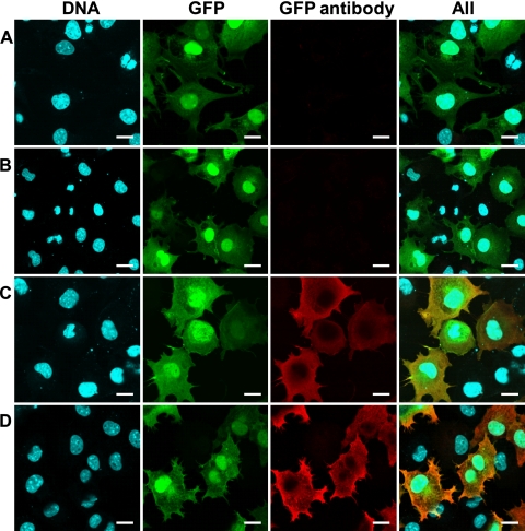
Fig 4.
Detection of antibodies against GFP in the sera of mice immunized with the PB2-KO virus. Confluent 293 cells that transiently express GFP were treated with sera (1/20 dilution) obtained from mice inoculated with medium (A), the formalin-inactivated virus (B), or the PB2-KO virus (C) or were treated with a commercial anti-GFP antibody (D). DNA (first column) was stained with Hoechst 33342. GFP (second column) represents cells transfected with a plasmid for the expression of GFP. GFP antibody (third column) represents the presence of the GFP antibody in the samples. These three images were merged (fourth column). Scale bars, 20 μm.

