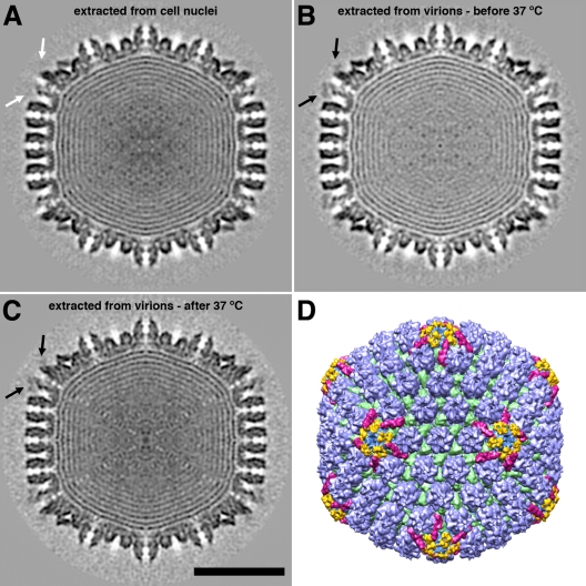
Fig 3.
Three-dimensional reconstructions of DNA-containing HSV-1 capsids. (A to C) Central sections through capsids viewed along a 2-fold axis of icosahedral symmetry. (A) Nuclear C-capsid; (B) T36 capsid isolated at 4°C; (C) T36 capsid isolated at 37°C. The black arrows in panels B and C point to additional density features overlying pentons. There is no such density feature in panel A (white arrow). Bar = 50 nm. (D) Color-coded surface rendering of the reconstruction shown in panel C. The surface features are shown in color as follows: capsomer protrusions are blue, triplexes are green, CCSCs are magenta, and the additional penton-associated density features (part of UL36) found on the T36 capsid are yellow.

