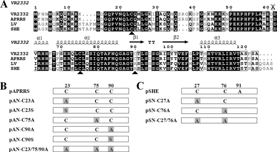
Fig 2.
Sequence alignment of PRRSV N proteins and the construction of cysteine mutants. (A) Alignment of N sequences from type II PRRSV strains (VR2332, GenBank accession no. U87392; APRRS, GQ330474) with the corresponding sequences from type I strains (LV, M96262; SHE, GQ461593). Completely conserved residues are indicated in black boxes, and partially conserved residues are in white boxes. The cysteines at positions 23, 75, and 90 of the type II N protein are indicated by black triangles. The scissors indicate the split site of the N protein for the BiFC assay. The secondary structure elements above the sequence show the structure of the C terminus of the VR2332 N protein (11). This figure was generated by ESPript (17), with slight modifications. (B) The cysteine residues in the N protein of APRRS and SHE were replaced with alanine or serine, either alone or in combination. The number above the boxes indicates the location of the cysteine or alanine. PCR-based site-directed mutagenesis was used to replace the codon for cysteine (TGC) with that of alanine (GCG) or serine (AGC).

