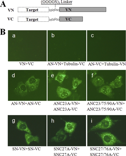
Fig 7.
BiFC analysis of full-length N proteins. (A) Schematic diagram of BiFC constructs. The Venus protein was split between amino acid residues 173 and 174, resulting in fragments VN (N-terminal 173 residues) and VC (C-terminal 66 residues). The so-called Target protein was fused to the N terminus of VN or VC with a linker sequence to generate the BiFC pair. (B) Visualization of interactions between full-length N proteins in living cells. Fragments VN and VC or α-tubulin gene-fused VN and VC were coexpressed in MARC-145 cells, and the fluorescent signals were examined as negative controls to assess the specificity of the BiFC method (panels a to c). Full-length N proteins from both genotypes of PRRSV with or without cysteine mutations were fused to the N terminus of VN or VC, and cells were cotransfected and examined for fluorescence (panels d to i).

