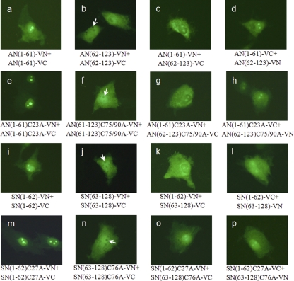
Fig 8.
BiFC analysis of truncated N proteins. Full-length N proteins with or without cysteine mutations were split between amino acid residues 61 and 62 for APRRS or between 62 and 63 for SHE. The resulting halves were ligated to VN or VC, MARC-145 cells were transfected, and fluorescent signals were visualized. The arrows indicate the nucleoli with no fluorescence.

