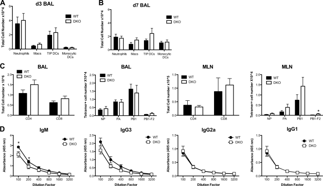
Fig 3.
Characterization of immune responses in WT and DKO mice. (A and B) Mice were infected intranasally with 2,000 EID50 of PR8 (WT mice, n = 15; DKO mice, n = 16). BAL fluid was collected at d3 (A) and d7 (B) postinfection; cells were analyzed by FACS; and cell numbers for neutrophils (CD11b+, Gr1+, major histocompatibility complex class II negative), macrophages (Macs; CD11b+, Gr1+, major histocompatibility complex class II positive), TNF-iNOS-producing DCs (TIP DCs; Ly6Chigh, CD11b+, CD11c+), and monocytic DCs (CD11b+, CD11c+) were enumerated. (C) Mice were infected intranasally with 2,000 EID50 of PR8, and BAL fluid was collected at d7 postinfection (WT mice, n = 18; DKO mice, n = 15). Cells from BAL fluid and MLN were analyzed by FACS, and total numbers of CD4+ T cells, CD8+ CTLs, and tetramer-positive CTLs were enumerated. *, P < 0.05, Student's t test. (D) PR8-specific serum antibody titers at d7 postinfection (WT mice, n = 11; DKO mice, n = 9) were determined by ELISA. *, P < 0.02, two-way analysis of variance. Data in panels A to C are from three independent experiments (mean and SEM).

