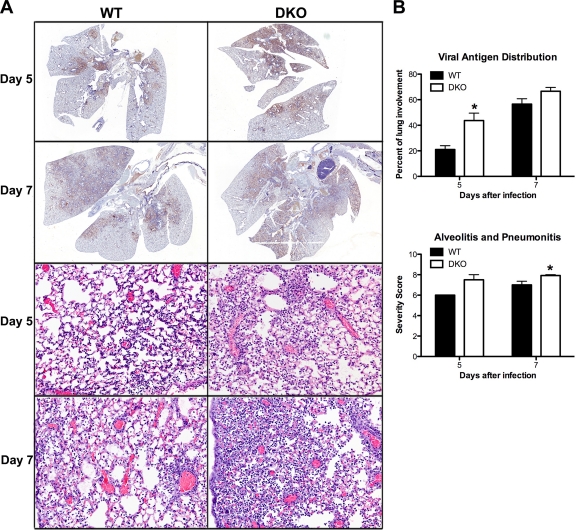
Fig 4.
Lung histology of flu-infected WT and DKO mice. WT and DKO mice were infected intranasally with 2,000 EID50 of PR8. Lungs were harvested for histopathological analysis at d5 (WT mice, n = 5; DKO mice, n = 8) and d7 (WT mice, n = 19; DKO mice, n = 14) postinfection. (A) Lung sections from d5 and d7 postinfection were immunohistochemically stained for flu antigen (top four panels, brown). Representative micrographs are shown. Magnification, ×5. Lung sections from d5 and d7 were stained with hematoxylin-eosin (bottom four panels). Representative micrographs are shown. Magnification, 200×. (B) Sections were scored blindly for flu antigen distribution (% involvement; top four panels) and the combined score for severity of alveolitis and interstitial pneumonitis (bottom four panels). Severity scoring criteria for lung lesions is the following: 0, no lesions; 1, minimal, focal to multifocal, inconspicuous; 2, mild, multifocal, conspicuous; 3, moderate, multifocal, prominent; 4, marked, diffuse or coalescing, lobar; 5, severe, diffuse consolidation, multilobar.

