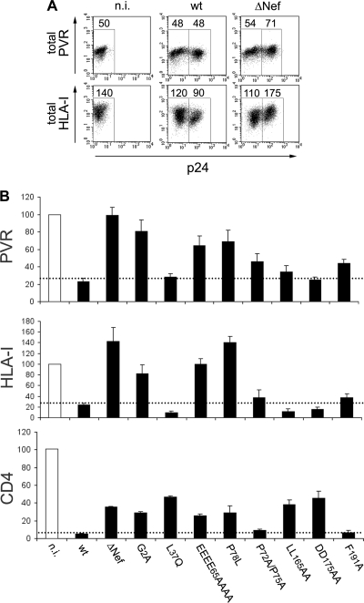
Fig 3.
Mechanisms of PVR modulation by HIV-1 Nef. (A) At 3 days postinfection, noninfected (n.i.) and wt- and ΔNef virus-infected Jurkat cells were permeabilized and then analyzed by two-color flow cytometry for total PVR or HLA-I levels as well as for p24 expression. Mean fluorescence intensity values are indicated. (B) Jurkat cells were not infected or infected with HIV-1 wt, ΔNef, or expressing a mutated Nef protein, as indicated, and then analyzed 3 days later for the expression of intracellular p24 and surface PVR or HLA-I, as described in the legend to Fig. 1A. Surface CD4 was analyzed at 1 day p.i. to limit the contribution of the late viral proteins Vpu and Env to the total CD4 downregulation. Surface expression of each molecule in cells infected with the indicated viruses was calculated by setting the mean fluorescence intensity obtained for n.i. cells to 100%. Values are averages ± SEs derived from at least three independent experiments. The dotted lines indicate the maximal surface expression found on wt HIV-infected cells.

