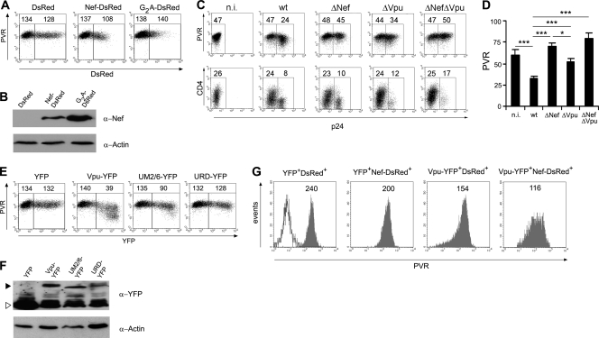
Fig 4.
Vpu contributes to the activity of Nef on surface PVR downregulation. (A) 293T cells were transfected with 0.5 μg of vector expressing DsRed, Nef-DsRed, or G2A-DsRed and analyzed 48 h later by two-color flow cytometry to measure DsRed fluorescence and cell surface PVR. Mean fluorescence intensity values relative to the value for PVR in DsRed-negative (left gate) and DsRed-positive (right gate) cells are indicated. (B) Total lysates of cells described in panel A were analyzed by Western blotting with anti-Nef and antiactin antibodies. (C) Jurkat cells were infected with HIV-1 either wt or defective for the expression of Nef, Vpu, or both proteins, as indicated, and analyzed for the surface expression of PVR or CD4 (on day 3 or 1 p.i., respectively), together with intracellular p24 accumulation. (D) The mean fluorescence intensity ± SE for PVR in n.i. and in p24+ infected cells was determined as described for panel C in seven independent experiments. *, P < 0.05; ***, P < 0.001. (E and F) 293T cells were transfected and analyzed as described for panels A and B, with the difference that the indicated YFP, Vpu-YFP, UM2/6-YFP, or URD-YFP proteins were expressed and monitored by YFP-specific fluorescence and anti-GFP/YFP Western blotting. The YFP-tagged Vpu proteins and YFP alone are indicated with a filled arrowhead and an open arrowhead, respectively. (G) 293T cells were doubly transfected with 0.5 μg of each vector in order to express DsRed or DsRed-Nef together with YFP or Vpu-YFP and then analyzed 48 h later by FACS. The expression of PVR on gated double-positive cells is indicated. The open histogram shows unlabeled cells. Results obtained with 293T cells (A, B, and E to G) are from one representative experiment out of three.

