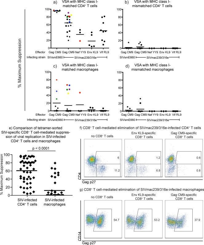
Fig 1.
Ex vivo tetramer-sorted SIV-specific CD8+ T cells suppressed viral replication in SIV-infected CD4+ T cells but were ineffective at suppressing viral replication in SIV-infected macrophages. We calculated the maximum percentage of viral suppression for each ex vivo tetramer-sorted SIV-specific CD8+ T cell population using the number of viral RNA (vRNA) copies per milliliter of culture supernatant at 48 h with and without effector cells: (vRNA copies/ml without CD8+ T cells − vRNA copies/ml with CD8+ T cells)/vRNA copies/ml without CD8+ T cells × 100. We used only tetramer-sorted SIV-specific CD8+ T cells that were greater than 50% specific as measured by postsort tetramer stains. The purity of the tetramer-sorted SIV-specific CD8+ T cells did not correlate with their ability to suppress viral replication in SIV-infected CD4+ T cells or macrophages. Experiments in panels a and c as well as panels b and d were directly matched: targets were derived from the same animals on the same day, infected simultaneously the same way 4 days after harvesting, and incubated with the same tetramer-sorted effectors for 48 h. We used nonautologous targets because effector cells were harvested from SIV-infected animals and using autologous targets would not allow for an MHC class I-mismatch control. (a) Ex vivo tetramer-sorted SIV-specific CD8+ T cells effectively suppressed viral replication in SIVmac239-, SIVmac239/316e-, and SIVsmE660-infected MHC class I-matched CD4+ T cell targets at an effector-to-target ratio of 1:1 after 48 h of coincubation. (b) Percent maximum suppression of SIV-specific CD8+ T cells incubated with MHC class I-mismatched SIV-infected CD4+ T cells. The range of viral replication in the SIV-infected CD4+ T cells without CD8+ T cells was 1 × 10⁶ to 1 × 10⁷/ml of viral RNA copies/ml of supernatant. (c) Ex vivo tetramer-sorted SIV-specific CD8+ T cells poorly suppressed both SIVmac239/316e- and SIVsmE660-infected MHC class I-matched macrophages. (d) Percent maximum suppression of SIV-specific CD8+ T cells incubated with MHC class I-mismatched SIV-infected macrophages. The range of viral replication in the SIV-infected macrophages without CD8+ T cells was 1 × 10⁵ to 1 × 10⁶ viral RNA copies/ml of supernatant. The average percent maximum suppression capacity is indicated for each animal with black bars. SIV-specific CD8+ T cell populations isolated from elite controllers are indicated with circles, while SIV-specific CD8+ T cell populations isolated from progressors are indicated with diamonds. The colored symbols in panel a correspond to the tetramer-sorted SIV-specific CD8+ T cell populations that suppressed viral replication in SIV-infected macrophages in panel c. Each data point represents the average of one experiment performed in duplicate or triplicate. Ex vivo tetramer-sorted SIV-specific CD8+ T cells were harvested from several time points throughout the chronic phase of infection of SIV-infected rhesus macaques. (e) Statistical comparison of all tetramer-sorted SIV-specific CD8+ T cell-mediated suppression of viral replication in SIV-infected CD4+ T cells and macrophages. The difference in suppression of viral replication observed between CD4+ T cells and macrophages was statistically significant (P < 0.0001). (f) Intracellular Gag p27 staining of a representative experiment of MHC class I-matched SIVmac239/316e-infected CD4+ T cells incubated for 48 h alone (left panel), with Mamu-B*08+ EnvKL9-specific CD8+ T cells (middle panel), or with Mamu-A*01+ GagCM9-specific CD8+ T cells (right panel). Dot plots were generated by gating on live, CD8− cells. (g) Intracellular Gag p27 staining of a representative experiment of MHC class I-matched SIVmac239/316e-infected macrophages incubated for 48 h alone (left panel), with Mamu-B*08+ EnvKL9-specific CD8+ T cells (middle panel), or with Mamu-A*01+ GagCM9-specific CD8+ T cells (right panel). Dot plots were generated by gating on live, HLA-DR+ CD14+ macrophages.

