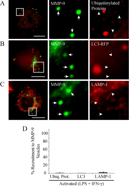
FIGURE 5.
MMP-9 vesicles do not colocalize with markers of proteosomal, lysosomal, or autophagic degradation. Some RAW 264.7 cells were transfected with RFP-tagged LC3. Cells were stimulated with LPS/IFN-γ (0.1 μg/ml and 100 units/ml, respectively, for 9 h). Cells were fixed and immunostained for MMP-9 (green) and immunostained for ubiquitinylated proteins (A) or analyzed for LC3-RFP (B) or immunostained for LAMP-1 (C) (red) and examined by epifluorescent microscopy. The arrows identify MMP-9 vesicles, whereas the arrowheads indicate the absence of co-recruitment to MMP-9 vesicles. These cells had a robust amount of MMP-9 vesicles and represent the relative degrees of colocalization with the indicated markers. Scale bars, 10 μm. The percentage of MMP-9 vesicles showing co-recruitment with the indicated markers was quantified (D). Error bars, S.E.

