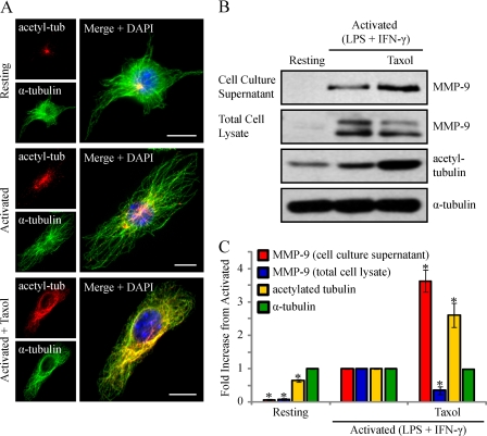
FIGURE 8.
Activated macrophages treated with taxol to enhance MT stabilization show increased levels of MMP-9 secretion. RAW 264.7 cells were activated (0.1 μg/ml LPS and 100 units/ml IFN-γ for 9 h) and treated with 0.1 μm taxol for 1 h. Cells were fixed and immunostained for acetylated α-tubulin (red), α-tubulin (green), and DAPI (blue) (A). Scale bars, 10 μm. Alternatively, the cell culture supernatants were collected, the cells were lysed, and the supernatants and lysates were subjected to Western blotting (B) and densitometric analysis (C) for MMP-9, acetylated tubulin, and α-tubulin. *, p < 0.05 compared with activated cells. Error bars, S.E.

