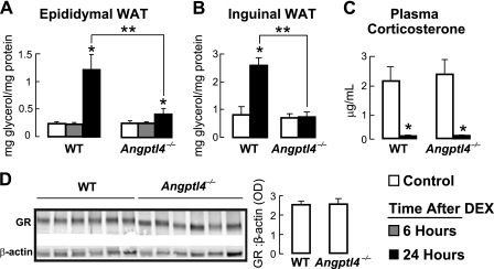
FIGURE 4.
A, the concentration of glycerol released into the medium over 2 h from epididymal WAT explants taken from control (PBS-treated) mice and from mice either 6 or 24 h following a single 5 mg/kg intraperitoneal dose of DEX, showing a reduction in the concentration of glycerol released by Angptl4−/− WAT 24 h after DEX treatment (n = 7–8; *, p < 0.05 versus control; **, p < 0.05 versus WT 24-h DEX). B, as for A, using inguinal WAT explants (n = 7–8; *, p < 0.0001 versus WT control; **, p < 0.001 versus WT 24-h DEX). C, plasma corticosterone levels, showing DEX-induced suppression across genotypes (n = 5–6; *, p < 0.05 versus control). D, WAT GR protein abundance by immunoblot (n = 6). The image is a grouping of representative images from different areas of the same gel. Error bars, S.E.

