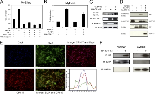
FIGURE 4.
PP1α-induced repression of myocardin is attenuated by CPI-17. A, COS7 cells were transfected with the myocardin enhancer (MyE), MEF2C, PP1α (PP1), activated RhoA (RhoA L63), or activated MKK6 and p38 (MKK6EE/p38), as indicated. Extracts were subjected to lucifierase assay. B, cells were transfected with the myocardin enhancer, and MEF2C, PP1, or CPI-17 as indicated, followed by luciferase assay. C, COS7 cells were transfected with HA-CPI-17 and HA-PP1α. Protein extracts were immunoprecipitated with PP1α antibody and immunoblotted, as indicated. D, COS7 cells were transfected with MEF2C, HA-PP1, or HA-CPI-17 as indicated. Extracts were immunoprecipitated with MEF2C antibody and immunoblotted for antibodies to HA. Protein extracts were immunoblotted, as indicated, to demonstrate equal loading and transfection efficiency. E, primary VSMCs were fixed, permeabilized, and subjected to immunofluorescence for CPI-17, smooth muscle actin (SMA), and the DAPI nuclear stain. Cells were visualized by standard fluorescence techniques. Relative fluorescence of a representative cell was graphed with ImageJ. F, A10 cells were transfected with HA-CPI-17 and subjected to nuclear/cytosolic fractionation. Lysates were immunoblotted as indicated. Error bars indicate S.E.

