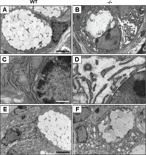
FIGURE 3.
Electron microscopy of goblet cells in the large intestine. A and B, images of goblet cells at the crypt base in the large intestine. C and D, high magnification of the rough ER in A and B. Note that rough ER in Oasis−/− goblet cells displays aberrant expansion. E and F, goblet cells in the upper portion of the crypt. The numbers of mucus vesicles are decreased, and the membrane of some vesicles are often fused in goblet cells of Oasis−/− large intestine. Scale bars = 4 μm (A, B, E, and F), 1.6 μm (C and D)

