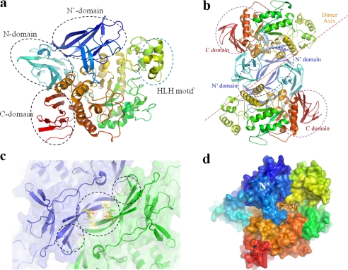
FIGURE 1.
Overall SMMA structure. a, a schematic overview of an SMMA monomer that shows the conserved N, catalytic, and C domains in CD-hydrolyzing enzymes with a novel N′ domain. The monomer is colored in a spectrum; the N terminus is in blue, and the C terminus is in red. b, the SMMA dimer structure is shown with a 2-fold axis perpendicular to the plane. c, the dimeric interface between the N′ domains is shown with the adjacent hydrophobic and charged interactions. d, a surface diagram of the monomer with a hypothetical cyclodextrin molecule (orange) is generated by superposition with the binary complex structure (Protein Data Bank entry 1GVI) to highlight the active site.

