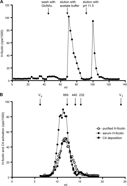
FIGURE 5.
Purification of H-ficolin from serum. A, purification of H-ficolin on an AcHSA column. A 4–8% PEG cut from serum was run through an HSA-coupled Sepharose column in TBS/tw/Ca2+, and the effluent was loaded onto an AcHSA-Sepharose column. The column was washed with GlcNAc to elute any remaining L-ficolin and H-ficolin and was subsequently eluted as follows: first with 1 m sodium acetate, pH 7.5, and next with 10 mm diethylamine, pH 11.5. B, H-ficolin containing fractions from A were pooled and concentrated and then subjected to GPC on a Superose 6 column. A serum sample was subjected to GPC on the same GPC column. The amount of H-ficolin in each fraction was quantified by TRIFMA, and was analyzed for the ability to activate and deposit C4 fragments onto an AcBSA surface. The elution volume of thyroglobulin (669 kDa), ferritin (440 kDa), and catalase (232 kDa) as well as the void volume (V0) and the total volume (VT) are given at top of figure.

