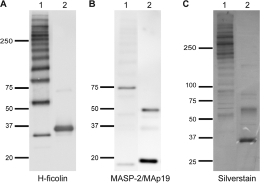
FIGURE 6.
Analysis of purified H-ficolin by SDS-PAGE and Western blotting. A, purified H-ficolin sample (pool from GPC, Fig. 5B) was run under nonreducing (lane 1) and reducing (lane 2) conditions, and the blot was developed with anti-H-ficolin antibody. The positions of the molecular mass markers (kDa) are indicated. B, same blot as in A developed with an anti-MASP-2/MAp19 antibody. C, H-ficolin containing fractions from the GPC were pooled and analyzed by SDS-PAGE and silver staining. Lane 1 was run under nonreducing conditions and lane 2 in reducing conditions.

