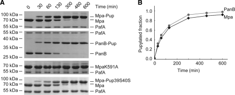
FIGURE 1.
Mpa is pupylated predominantly on a single lysine residue, and pupylation occurs on a time scale similar to that for the proteasomal substrate PanB. A, Pup conjugation to Mpa (1 μm hexamer, gel), PanB (6 μm monomer, second gel), the pupylation site variant MpaK591A (1 μm hexamer, third gel), or Mpa (1 μm hexamer) with the Pup39S40S variant (fourth gel) by PafA (1 μm or 5.25 μm for the Pup variant) visualized at various time points by SDS-PAGE and Coomassie staining. B, analysis of Mpa wild-type and PanB pupylation time trace by gel band densitometry (A, first and second gels).

