FIGURE 1.
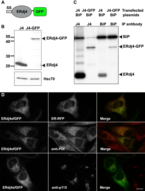
ERdj4-GFP behaves like the untagged ERdj4. A, schematic illustration of the ERdj4-GFP fusion construct. B, immunoblot of COS-1 cells transiently transfected with ERdj4 or ERdj4-GFP and lysates were subjected to immunoblotting with anti-ERdj4. Anti-Hsc70 staining indicates that similar amounts of cell lysates were loaded. C, immunoprecipitated ERdj4-GFP and ERdj4 interact with BiP in vivo. COS-1 cells were transiently transfected with plasmids expressing ERdj4 or ERdj4-GFP in combination with BiP. The cells were labeled 24 h post-transfection with [35S]methionine for 1 h, and the proteins were cross-linked by treating with 150 μg/ml dithiobis(succinimidyl propionate). The cell lysates were immunoprecipitated with either an anti-ERdj4 or anti-BiP antibody, as indicated. D, co-localization of ERdj4sfGFP with ER-RFP and anti-protein disulfide isomerase (PDI) by immunofluorescence staining, but not with the Golgi complex marker, p115. Scale bar, 10 μm.

