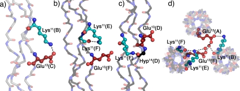
FIGURE 2.
Interstrand and interhelical interactions in KGE. a–c, interstrand hydrogen bonds involving the charged side chains. d, top view of the packing interactions involving the residues depicted in b. Triple helices oriented N to C terminus in Fig. 1 are shown in gray (A, leading chain; B, middle chain; C, lagging chain), and triple helices oriented C to N terminus in Fig. 1 are depicted in black (D, leading chain; E, middle chain; F, lagging chain). Lysines are shown in cyan and glutamates in red. Amino acids are labeled using their three-letter code, sequence position, and chain. Images were generated using PyMOL (21).

