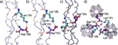
FIGURE 3.
Interstrand, intrastrand, and interhelical interactions in KGD. a and b, interstrand hydrogen bonds involving the charged side chains. c, intrastrand hydrogen bond. d, top view of packing interactions involving the residues depicted in b and c. Triple helices oriented N to C terminus in Fig. 1 are shown in gray (A, leading chain; B, middle chain; C, lagging chain), and triple helices oriented C to N terminus in Fig. 1 are depicted in black (D, leading chain; E, middle chain; F, lagging chain). Lysines are shown in cyan and aspartates in purple. Amino acids are labeled using their three-letter code, sequence position, and chain. Images were generated using PyMOL (21).

