FIGURE 1.
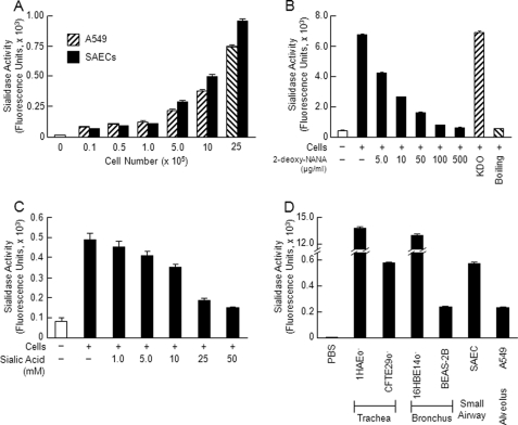
EC sialidase activity. A, increasing SAEC and A549 cell numbers were assayed for sialidase activity for the fluorogenic substrate 4-MU-NANA. B, A549 cells (1.0 × 10⁶) were assayed for sialidase activity for 4-MU-NANA prior to and after boiling and in the presence of increasing concentrations of 2-deoxy-NANA (5–500 μg/ml) or its negative control, 2-keto-3-deoxyoctulosonic acid (KDO) (500 μg/ml). C, A549 cells (1.0 × 10⁶) were assayed for 4-MU-NANA in the presence of increasing concentrations of N-acetylneuraminic acid (1–50 mm). D, equal numbers of airway ECs (1.0 × 10⁶) derived from the trachea (1HAEo− and CFTE29o−), bronchus (16HBE14o− and BEAS-2B), small airways (SAECs), and alveolus (A549) or PBS as a negative control was assayed for sialidase activity for the 4-MU-NANA substrate. Vertical and error bars represent mean ± S.E. sialidase activity (n ≥ 2) expressed as arbitrary fluorescence units. The results in each panel represent ≥2 independent experiments.

