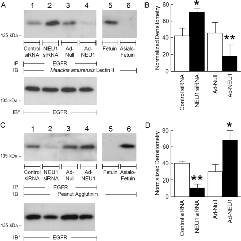
FIGURE 4.
EGFR is NEU1 substrate in airway ECs. A549 cells were transfected with NEU1-targeting or control siRNAs or infected with Ad-NEU1 or Ad-Null at an m.o.i. of 100. Cell lysates were immunoprecipitated with anti-EGFR antibody. The EGFR immunoprecipitates were resolved by SDS-PAGE and probed with MAL (A) or PNA (C). As controls for lectin specificity, parallel blots of fetuin and asialofetuin were simultaneously probed (lanes 5 and 6). To control for protein loading and transfer, blots were stripped and reprobed with anti-EGFR antibody. IP, immunoprecipitation; IB, immunoblot; IB*, immunoblot after stripping. Molecular mass in kDa is indicated on the left. Each blot is representative of two independent experiments. The densitometric analyses of the blots in A and C are presented in B and D, respectively. Vertical and error bars represent mean ± S.E. MAL/PNA signal normalized to EGFR signal in the same lane on the same blot (n = 2). *, significantly increased lectin/EGFR densitometry of NEU1 siRNA- or Ad-NEU1-treated cells compared with control siRNA- or Ad-Null-treated cells at p < 0.05. **, significantly decreased lectin/EGFR densitometry of NEU1 siRNA- or Ad-NEU1-treated cells compared with control siRNA- or Ad-Null-treated cells at p < 0.05.

