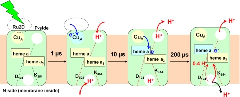
FIGURE 4.
Model for coupling electron injection and proton transfer steps in O → E step. A nanosecond laser flash excites Ru2D and initiates electron transfer to CuA within 1 μs. This event causes electron uptake from both sides of the enzyme. Red arrows indicate proton movement in the direction of proton pumping through COX, and black arrows indicate proton transfer in the opposite direction. After 10 μs, the electron is being transferred to heme a, and partial proton release occurs on the P-side of the membrane. After 200 μs, all but 0.38 protons/COX are released. According to Ref. 23, the residual proton is transferred toward the catalytic center.

