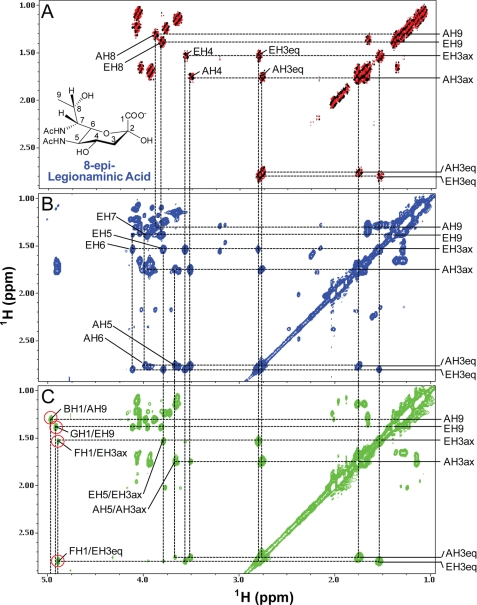
FIGURE 10.
DQF-COSY (A), TOCSY (B), and NOESY (C) spectra of the wild-type V. fischeri O-antigen oligosaccharide. These data illustrate the assignments of the spin systems for the sugar residues A and E of 8-epi-legionaminic acids. The DQF-COSY spectrum (A) is shown in black and red, and the TOCSY (B) and NOESY (C) spectra are shown in blue and green, respectively. Some of the diagonal peaks of residues A and E are labeled along the right side of the panels. Some of the off-diagonal cross-peaks of the residues A and E are also labeled. The 1st letter in the labels refers to sugar subunit and the rest in the labels refers to the position on that sugar residue. The inter-glycosidic NOESY cross-peaks are indicated by red circles in C. The TOCSY spectrum was collected with a mixing time of 103 ms, and the NOESY spectrum was acquired with a mixing time of 160 ms. The structure and position labeling of the 8-epi-legionaminic acid are indicated in A.

