FIGURE 2.
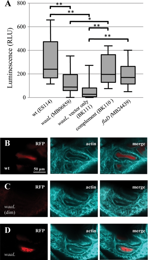
Animal colonization by waaL mutant. A, box-whisker plot of luminescence, in relative luminescence units (RLU), of animals colonized with different bacterial strains 24 h into exposure. n = 10 animals for each treatment except n = 20 for MB06859. Statistical comparison of treatments is by Mann-Whitney U test, following Kruskal-Wallis test for all data. Any two treatments not marked with an asterisk were not found to be significantly different (*, p < 0.05; **, p < 0.01). B–D, confocal microscopy of light organ crypts (actin cytoskeleton, shown in teal) in animals colonized with red fluorescent protein-labeled V. fischeri (red) at 24 h into exposure. B shows an animal exposed to wild-type bacteria, and C and D show animals exposed to the waaL mutant. The animal in C had no appreciable luminescence (dim) at the time of fixation (defined as relative luminescence units <2), but the animal in D was noticeably luminescent.

