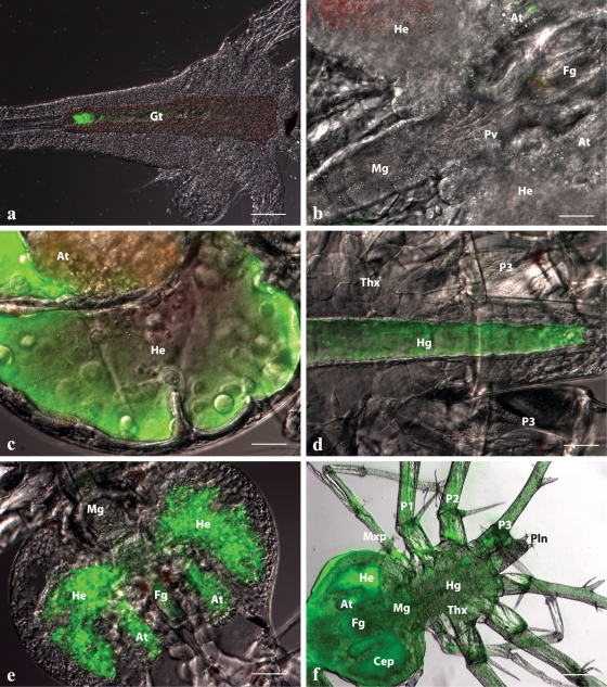
Fig 3.
In situ spatiotemporal localization of V. owensii DY05[GFP] in Artemia nauplius after 2-h enrichment (a) and during the infection process of vector-challenged stage 1 P. ornatus phyllosomas (b to f). (a) Artemia nauplius showing colonization of DY05[GFP] in the gastrointestinal tract. Scale bar, 100 μm. (b) Hepatopancreas and midgut region of phyllosoma after 6-h exposure, demonstrating the presence of single cells or small aggregations of DY05[GFP] cells in the foregut, the midgut, and the anterior and lateral lobes of the hepatopancreas. Scale bar, 50 μm. (c) Proliferation of DY05[GFP] in the distal ends of the lateral and anterior hepatopancreas lobes 12 h after exposure. Scale bar, 50 μm. (d) Hindgut 12 h after exposure showing trafficking of DY05[GFP] cells followed by evacuation. Scale bar, 50 μm. (e) Hepatopancreas 18 h after exposure, showing illumination of the entire organ with fluorescent DY05[GFP] cells concomitant with tissue granulation and loss of architecture. Scale bar, 100 μm. (f) Dead phyllosoma 24 h after exposure, showing colonization of the entire body (systemic infection) by DY05[GFP] cells associated with loss of internal organ structural integrity. Scale bar, 200 μm. At, hepatopancreas anterior lobe; Cep, cephalic shield; Fg, foregut; Gt, gastrointestinal tract; He, hepatopancreas lateral lobe; Hg, hindgut; Mg, midgut; Mxp, maxilliped; P, pereiopod (1 to 3); Pln, pleon; Pv, pyloric valve; Thx, thorax.

