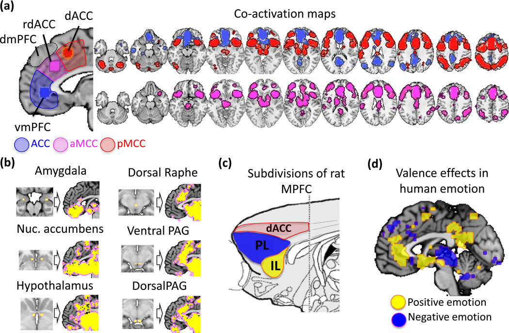
Figure I.
Subdivisions and connectivity of the medial prefrontal cortex. (a) Four functionally distinct zones include ventromedial prefrontal cortex (vmPFC), rostral dorsal anterior cingulate (rdACC), dorsal anterior cingulate (dACC), and dorsomedial prefrontal cortex (dmPFC). We envision meaning construction as occurring principally in vmPFC and rdACC, with substantial contributions from dmPFC. To illustrate the differential connectivity of the vmPFC and dACC zones, ‘seed’ regions were selected based on the peak activation frequencies across the 1,669 studies and 8 domain areas summarized in this review. Areas significantly co-activated with each seed region are shown at the right (see Supplementary Online Materials for details). The dACC (red) and vmPFC (blue) regions co-activated with distinct brain networks. Additional subcortical connectivity with vmPFC was apparent below the stringent thresholds used here (P < .05 corrected). rdACC (purple) showed co-activation with both networks, and particularly strong co-activation with periaqueductal gray (PAG) and other subcortical areas. (b) Further exploration of medial prefrontal co-activation with subcortical regions. For each of the six areas shown, small ‘seed’ regions were placed in the subcortical area, and co-activated areas in the medial prefrontal cortex are shown (P < .0001). rdACC is co-activated with each area, with particularly strong co-activation with dorsal PAG and raphe nuclei associated with active responses to threat, suggesting a possible homology with prelimbic cortex in rats. (c) Location of prelimbic and infralimbic cortices in rats, adapted from [99]. (d) A meta-analytic summary of 138 PET and fMRI studies of positive and negative emotional experience (374 experimental contrasts, selected from a database of 234 studies of emotion described in [88]). Colored regions indicate areas with significantly greater density of activations related to positive (yellow) and negative (blue) emotional experience. Positive emotions more consistently activate the vmPFC, posterior cingulate cortex, ventral striatum and supplementary motor areas, and pre-supplementary motor area. Negative emotions more consistently activate the PAG, rdACC, dmPFC and deep cerebellar areas.

