
Figure 1. High-resolution computed tomographic images demonstrating bronchial wall thickening.
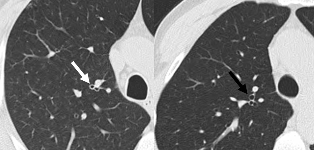
Images are from similar lung anatomic levels from a rheumatoid arthritis (RA)-related autoantibody positive case (left image; anti-cyclic citrullinated peptide antibody and rheumatoid factor isotypes IgM and IgA positive), and a RA-related autoantibody negative control (right image) that was matched to the case on age, sex and smoking status. The left image demonstrates bronchial wall thickening (white arrow). In contrast, the right image demonstrates a normal-appearing thin-walled bronchus (black arrow).