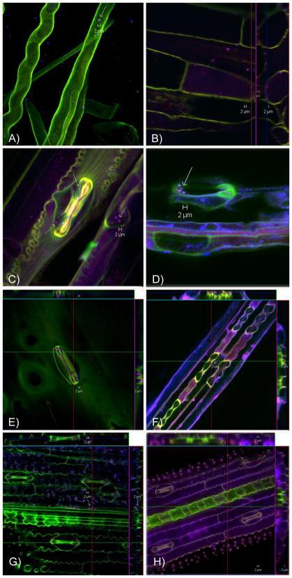
Figure 2.
FISH and confocal laser-scanning microscopy on roots and leaves using a validated set of fluorescently labeled oligonucleotides targeting the 16S rRNA of B. pseudomallei (red); β-Proteobacteria (blue) as positive control and the probe mix non-EUB-338-I, II, III (green) as negative control for nonspecific probe or dye binding. The composite pictures are shown with B. pseudomallei cells in magenta (combination of red and blue). No unspecific green signals were evident for B. pseudomallei signals (see Supplement Figure 3). The orthogonal views (B, E, F, G, H) depict the intra- or extracellular location of B. pseudomallei by showing an internal of three dimensional z-stacks. A) Two B. pseudomallei are seen within a root hair (inside location confirmed by orthogonal view – see Supplement Figure 3) and further β-Proteobacteria among root hairs of Tully Grass whose rhizosphere was inoculated with B. pseudomallei. B) B. pseudomallei in the rhizosphere and inside root cells of Mission Grass from a highly positive field site. C) Various B. pseudomallei and other β-Proteobacteria are shown close to and within stomatal guard cells of a Mission Grass leaf of a highly positive field site. D) Three B. pseudomallei outside of a stomatal opening of a leaf of an introduced pasture grass (Digitaria milanjiana cultivar Jarra) whose rhizosphere was inoculated with B. pseudomallei. E) B. pseudomallei in stomatal guard cells and on the surface of a stomata of a Paspalum leaf collected at field site B. F)–H) B. pseudomallei are seen along vascular bundles of leaves of grasses whose rhizosphere was inoculated with B. pseudomallei. They are at the surface of the lower side of the leaf in a gully-like structure created by the vascular bundles F) Leaf of Sorghum intrans. G)–H) Leaves of Wild Rice.

