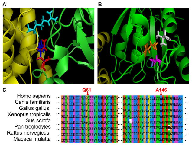
Figure 3. Illustration of two disease-causing mutations (Q61K and A146T) in human HRAS.
a) Structural representation of the Q61K mutation. Chains A and B are shown in green and yellow, respectively. The residue Gln 61 (wild-type) is shown in red, Lys 61 (mutant) in blue, and Arg 47 of chain B in cyan. The hydrogen bond is represented as a white dash line.
b) Structural representation of the A146T mutation. Chains A and B are shown in green and yellow, respectively. Ala 146 (wild-type) is shown in magenta, and Thr 146 (mutant) in blue. Two neighboring residues, Leu 15 in orange and Val 148 in white, are also shown.
c) Multiple sequence alignment of HRAS with the amino acid substitution sites indicated.

