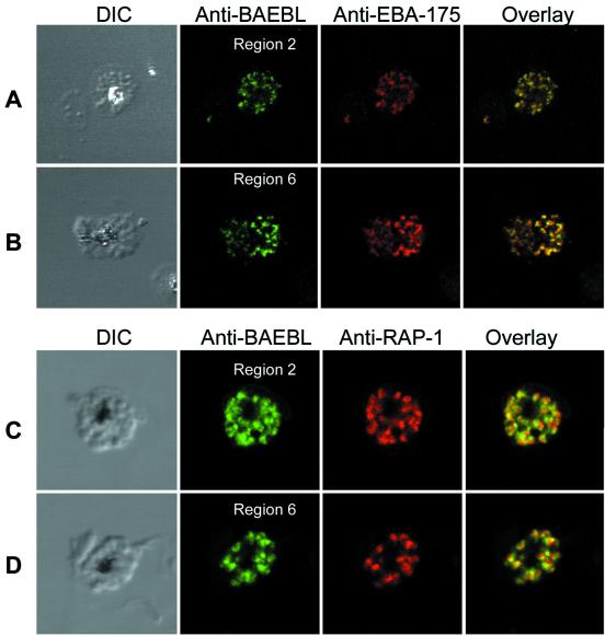
Figure 2.
Confocal microscopy demonstrates the localization of BAEBL in micronemes. (A) Dd2/Nm schizonts were double labeled with anti-BAEBL region 2 and anti-EBA-175. Schizonts immunolabeled with anti-BAEBL region 2 were stained with Alexa 488 secondary antibody (green). Schizonts labeled with anti-EBA-175 were stained with Alexa 594 secondary antibody (red). (B) Dd2/Nm schizonts were double labeled with anti-BAEBL region 6 and anti-EBA-175. Schizonts immunolabeled with anti-BAEBL region 6 were stained with Alexa 488 secondary antibody (green). Schizonts labeled with anti-EBA-175 were stained with Alexa 594 secondary antibody (red). (C) Dd2/Nm schizonts were double labeled with anti-BAEBL region 2 and anti-RAP-1 monoclonal antibody. Schizonts immunolabeled anti-BAEBL region 2 were stained with Alexa 488 secondary antibody (green). Schizonts labeled with anti-RAP-1 were stained with tetramethyl rhodamine isothiocyanate (TRITC) secondary antibody (red). (D) Dd2/Nm schizonts were double labeled with anti-BAEBL region 6 and anti-RAP-1 monoclonal antibody. Schizonts immunolabeled with anti-BAEBL region 6 were stained with Alexa 488 secondary antibody (green). Schizonts labeled with anti-RAP-1 were stained with TRITC secondary antibody (red).

