
Fig. 9.
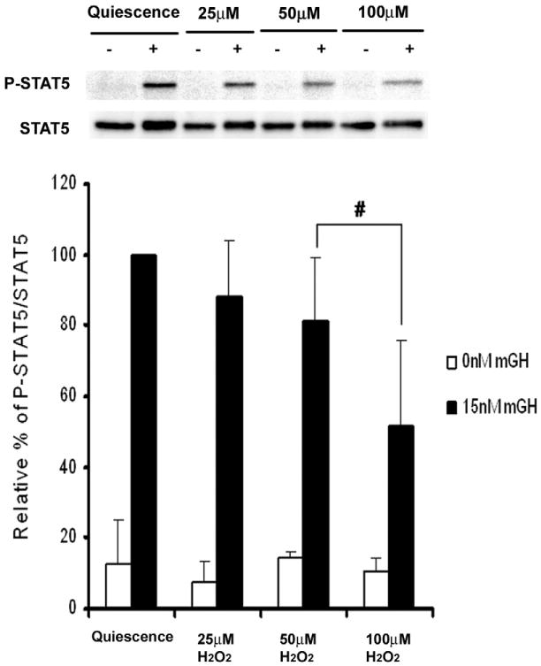
Effect of hydrogen peroxide on growth hormone-stimulated STAT5 tyrosine phosphorylation in βTC-6 cells. βTC-6 cells were incubated 21–22 h in quiescence medium and then incubated with 0, 25, 50, or 100 μM hydrogen peroxide for 30 min before cytokine stimulation. Cells were then stimulated for 15 min without or with 15 nM recombinant mouse growth hormone (mGH). The cells were then lysed, and STAT5 was immunoprecipitated from the PAS-precleared lysate and analyzed via SDS–PAGE and Western immunoblot as described under Material and methods to measure the ratio of phosphorylated STAT5 to total STAT5; these ratios were expressed as the mean (n = 3) ± standard deviation. #P<0.05 relative to mGH-stimulated, nonoxidized quiescent cells.