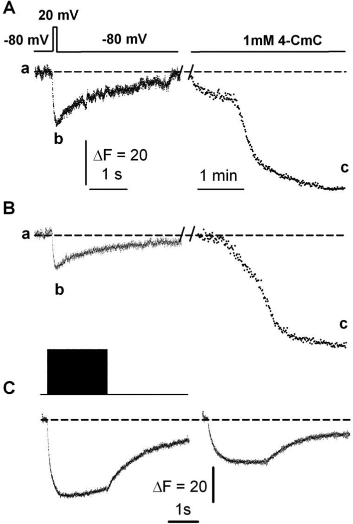
Figure 6. Maximal SR Ca2+ depletion evoked by 4-CmC.
A. CatchER fluorescence transient (bottom trace) in response to 20mV/100 ms command pulse (top trace) in a voltage-clamped myofiber from a young-adult mouse. The same fiber was exposed to 1mM 4-CmC with ~2min interval. The dashed line indicates the basal fluorescence (a), while the nadir of the response to electrical stimulation or 4-CmC application is shown in (b) and (c), respectively. B. CatchER fluorescence transient in response to a 20mV/100 ms command pulse in a voltage-clamped myofiber from an old mouse. The same fiber was exposed to 1mM 4-CmC at ~2min intervals. C. CatchER fluorescence (bottom) in response to repetitive stimulation with 2-ms pulses at 150 Hz for 2 s at 20mV (top) in myofibers from young (left) and old (right) mice.

