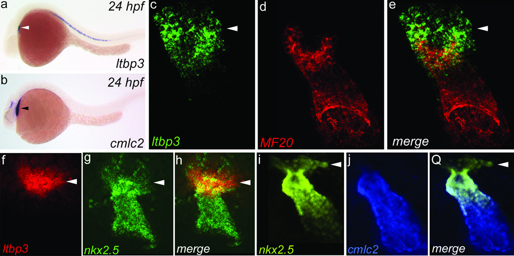
Figure 1. ltbp3 and nkx2.5 transcripts mark extra-cardiac cells contiguous to the outflow pole of the zebrafish heart tube.
a,b, ltbp3+ cells (white arrowhead) visualized by in situ hybridization at 24 hours post-fertilization (hpf) reside dorsal to the cmlc2+ heart tube (black arrowhead; n=15+ embryos/group). c–e, Heart tube region in 24 hpf embryo co-stained with ltbp3+ riboprobe (green; arrowhead) and a muscle-specific antibody (MF20; red; n=3/3) that recognizes cardiomyocytes. f–h, Heart tube region in 24 hpf embryo co-stained with ltbp3 and nkx2.5 riboprobes highlighting ltbp3+, nkx2.5+ cells (arrowheads) at the outflow pole of the heart tube (n=9/9). i–k, Heart tube region in 26 hpf Tg(nkx2.5::ZsYellow); Tg(cmlc2::CSY) embryo highlighting non-myocardial nkx2.5+ cells (arrowheads).

