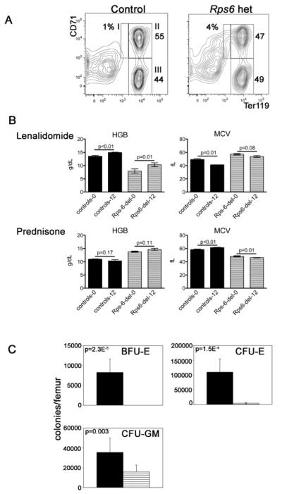
Figure 1. Hematologic characterization of Rps6 heterozygously-deleted mice.
(A) Representative flow cytometric analyses of whole bone marrow immunostained with antibodies to CD71 (transferrin receptor) and Ter119 (erythroid specific). The relative percentages of nucleated cells in each of the populations I to III are indicated. We found a statistically significant increase in the percentage of cells in population I in Rps6 heterozygously-deleted mice, 4.4%±1.0 vs. 1.2±0.1, p= 0.03, mean±SEM, two-tailed Student’s t-test, 4 mice in each group. Analyses of spleens showed similar findings. (B) Hemoglobin (HGB) and mean corpuscular values (MCV) of Rps6 heterozygously-deleted mice (striped, Rps-del-0 and Rps-del-12) and controls (solid, controls-0 and controls-12) treated with lenalidomide (top, rpS6 hets n= 7, controls n=5) or prednisone (bottom, rps6 hets n= 19, controls n= 8) for 12 weeks (baseline and 12-week values). The administered dose was based on pharmacokinetic studies in humans and rats[24] and communication with Celgene Corporation; it is estimated to achieve an ~2.2μM concentration of drug in the mice, equivalent to the therapeutic concentration achieved with a 25 mg/day dose of lenalidomide to patients with multiple myeloma[25]. Prednisone was dosed in their daily ad libitum food supply at approximately 2 mg prednisone/kg body weight per day (assuming a 20 gram mouse eating 4 grams diet/day). We excluded the possibility that the improvement in hemoglobin with lenalidomide reflects a preferential expansion of normal (undeleted) cells in the lenalidomide cohort by Southern blot (SK data not shown). A cohort of Rps6 heterozygously-deleted and control mice, age-matched to the animals treated with prednisone or lenalidomide were followed with monthly CBC analyses (n=3 in each group); in these animals the hemoglobin was not statistically different between the start and end of 12 weeks of monitoring (data not shown). We also analyzed the MCV and hemoglobin data in the prednisone-treated cohort as individual mice and observed no response to therapy. Our treatment studies are limited by not following drug levels in animals.
(C) Hematopoietic colony assays in Rps6 heterozygously-deleted mice (striped, n=6) and controls (solid, n=6). Mean±SEM, two-tailed Student’s t-test. That CFU-E were present while BFU-E were not detected suggests that our culture conditions did not optimally support BFU-E to CFU-E differentiation (since the detection of BFU-E in this assay requires that the plated BFU-E differentiate through the CFU-E-stage for enumeration), which has been reported in colony assays of DBA patients[26].

