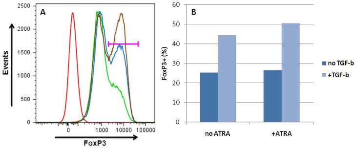
Figure 1. Human iTreg generation with TGF-β and ATRA.
Human CD4+ T cells were obtained by magnetic bead sorting (CD4+ kit, Miltenyi) of PBMC (purity >90%; 5% FoxP3+) and stimulated with high-dose IL-2 +/− TGF-β (20 ng/ml) +/− ATRA (10 ng/ml). After 6 days, live CD4+ cells were assessed for FoxP3 expression by flow cytometry. Cells cultured in the presence of TGF-β + ATRA showed 50% FoxP3+ expression while cells cultured with only IL-2 were only 25% FoxP3+; the remaining 75% of the cells showed an intermediate level of FoxP3.
(A) Gating for FoxP3+ cells (horizontal bar). Red: isotype; green: no TGF-β, no ATRA; blue: TGF-β only; brown: TGF-β + ATRA.
(B) Quantification of FoxP3+ cells: 50% of cells cultured with TGF-β + ATRA acquired FoxP3+ phenotype.

