Figure 1.
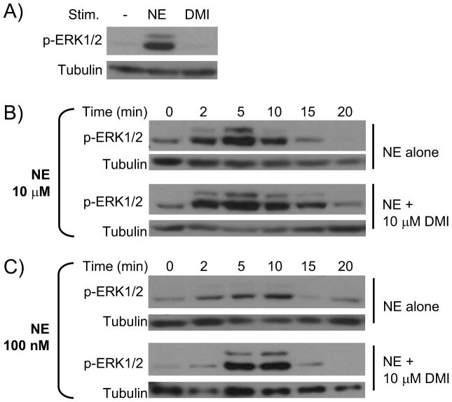
DMI impacts the kinetics of NE-induced α2AAR-mediated ERK1/2 signaling. All NE stimulations were done in the presence of propranolol (βAR antagonist) and prazosin (α1 and α2B/CAR antagonist). A. DMI does not itself drive ERK1/2 signaling. MEF cells were stimulated for 5 minutes with NE alone (as a positive control) or DMI alone. Whole cell homogenates were then analyzed by SDS-PAGE/Western blot, probing for phospho-ERK1/2 and tubulin (loading control). B. & C. MEF cells were stimulated for the indicated times with either 100 nM or 10 μM NE, alone or in combination with 10 μM DMI, and analyzed for ERK1/2 activation as in panel A. Blots shown are representative of 3 independent experiments.

