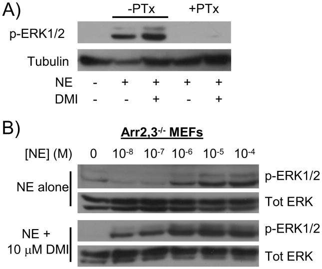
Figure 4.
DMI-modulated ERK1/2 signaling through the α2AAR remains Gi-dependent and arrestin-independent. A) MEF cells were subjected to pertussis toxin (+PTx) or vehicle (-PTx) treatment as described in methods, then stimulated with 10 μM NE alone or in combination with 10 μM DMI for 5 minutes. Whole cell homogenates were analyzed by SDS-PAGE/Western blot, probing for phospho-ERK1/2 and tubulin. B) Arrestin2,3 double knockout (Arr2,3−/−) MEFs were stimulated for 5 minutes with NE at varying concentrations, ranging from 10 nM (10−8 M) to 100 μM (10−4 M), alone or in combination with 10 μM DMI, as indicated. Homogenates were analyzed as in panel A, with the addition of stripping/probing for total ERK. Blots are representative of 3 independent experiments.

