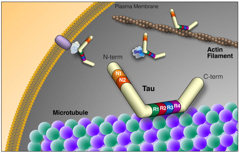
Figure 1. Tau Structure and Function.
Tau is an intrinsically disordered protein that can be alternatively spliced at N terminal exons (N1, 2) and the microtubule repeat domains (R). The domains of tau bind many different types of molecules, suggesting a central role in signaling pathways and cytoskeletal organization. The diversity of tau binding partners is highlighted in Table 1. N-term, N-terminus; C-term, C-terminus; SH3, protein SH3 domain

